Notes
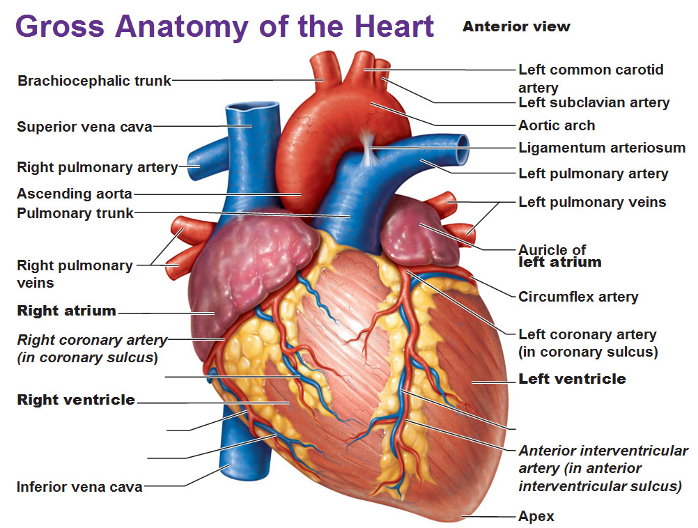
CARDIOGENIC SHOCK: PATHOPHYSIOLOGY
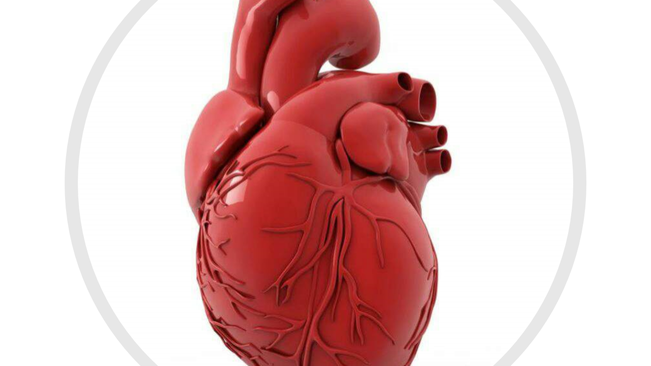
Cardiogenic shock also sometimes known as “pump failure” is a condition of diminished cardiac output that severely impairs cardiac perfusion. It reflects severe left-sided heart failure.
Pathophysiology
- Inability to contract. When the myocardium can’t contract sufficiently to maintain adequate cardiac output, stroke volume decreases and the heart can’t eject an adequate volume of blood with each contraction.
- Pulmonary congestion. The blood backs up behind the weakened left ventricle, increasing preload and causing pulmonary congestion.
- Compensation. In addition, to compensate for the drop in stroke volume, the heart rate increases in an attempt to maintain cardiac output.
- Diminished stroke volume. As a result of the diminished stroke volume, coronary artery perfusion and collateral blood flow is decreased.
- Increased workload. All of these mechanisms increase the heart’s workload and enhance left-sided heart failure.
- End result. The result is myocardial hypoxia, further decreased cardiac output, and triggering of compensatory mechanisms to prevent decompensation and death.
Classification
The causes of cardiogenic shock are known as either coronary or non-coronary.
- Coronary. Coronary cardiogenic shock is more common than noncoronary cardiogenic shock and is seen most often in patients with acute myocardial infarction.
- Noncoronary. Noncoronary cardiogenic shock is related to conditions that stress the myocardium as well as conditions that result in an ineffective myocardial function.
Causes
Cardiogenic shock can result from any condition that causes significant left ventricular dysfunction with reduced cardiac output.
- Myocardial infarction (MI)
- Myocardial ischemia
- End-stage cardiomyopathy
Clinical Manifestations
Cardiogenic shock produces symptoms of poor tissue perfusion.
- Clammy skin. The patient experiences cool, clammy skin as the blood could not circulate properly to the peripheries.
- Decreased systolic blood pressure. The systolic blood pressure decreases to 30 mmHg below baseline.
- Tachycardia. Tachycardia occurs because the heart pumps faster than normal to compensate for the decreased output all over the body.
- Rapid respirations. The patient experiences rapid, shallow respirations because there is not enough oxygen circulating in the body.
- Oliguria. An output of less than 20ml/hour is indicative of oliguria.
- Mental confusion. Insufficient oxygenated blood in the brain could gradually cause mental confusion and obtundation.
- Cyanosis. Cyanosis occurs because there is insufficient oxygenated blood that is being distributed to all body systems.
Assessment and Diagnostic Findings
Diagnosis of cardiogenic shock may include the following diagnostic tests:
- Auscultation. Auscultation may detect gallop rhythm, faint heart sounds and, possibly, if the shock results from rupture of the ventricular septum or papillary muscles, a holosystolic murmur.
- Pulmonary artery pressure (PAP). PAP monitoring may show increase in PAP, reflecting a rise in left ventricular end-diastolic pressure and increased resistance to the afterload.
- Arterial pressure monitoring. Invasive arterial pressure monitoring may indicate hypotension due to impaired ventricular ejection.
- ABG analysis. Arterial blood gas analysis may show metabolic acidosis and hypoxia.
- Electrocardiography. Electrocardiography may show possible evidence of acute MI, ischemia, or ventricular aneurysm.
- Echocardiography. Echocardiography can determine left ventricular function and reveal valvular abnormalities.
- Enzyme levels. Enzyme levels such as lactic dehydrogenase, creatine kinase. Aspartate aminotransferase and alanine aminotransferase may confirm MI.
Medical Management
The aim of treatment is to enhance cardiovascular status by:
- Oxygen. Oxygen is prescribed to minimize damage to muscles and organs.
- Angioplasty and stenting. A catheter is inserted into the blocked artery to open it up.
- Balloon pump. A balloon pump is inserted into the aorta to help blood flow and reduce workload of the heart.
- Pain control. In a patient that experiences chest pain, IV morphine is administered for pain relief.
- Hemodynamic monitoring. An arterial line is inserted to enable accurate and continuous monitoring of BP and provides a port from which to obtain frequent arterial blood samples.
- Fluid therapy. Administration of fluids must be monitored closely to detect signs of fluid overload.
Drug Treatment
Drug used may include:
- IV dopamine. Dopamine, a vasopressor, increases cardiac output, blood pressure, and renal blood flow.
- IV dobutamine. Dobutamine is an inotropic agent that increase myocardial contractility.
- Norepinephrine. Norepinephrine is a more potent vasoconstrictor that is taken when necessary.
- IV nitroprusside. Nitroprusside is a vasodilator that may be used with a vasopressor to further improve cardiac output by decreasing peripheral vascular resistance and reducing preload.
Source: Nurseslabs
Discover more from Nursing In Ghana
Subscribe to get the latest posts sent to your email.


Hypotension, commonly referred to as “low blood pressure,” is a medical condition in which the blood pressure in the arteries is lower than normal (when the blood pressure reading is lower than 90/60mmHg). There are various types of hypotension, each with different causes, symptoms, and treatments. As a nurse, it is important to be aware of the different types of hypotension and their management in order to provide safe and effective care to your patients.
Orthostatic hypotension
Orthostatic hypotension is a type of hypotension that occurs when a person changes position from lying down or sitting to standing up. This can cause a sudden drop in blood pressure, leading to symptoms such as dizziness, lightheadedness, and fainting. Orthostatic hypotension is common in older adults, especially those with underlying medical conditions such as Parkinson’s disease, diabetes, or autonomic neuropathy.
The management of orthostatic hypotension involves lifestyle modifications, such as avoiding sudden changes in position, staying hydrated, and wearing compression stockings. Medications such as fludrocortisone, midodrine, and droxidopa may also be prescribed to help raise blood pressure.
Neurally mediated hypotension.
Neurally mediated hypotension also known as the fainting reflex, neurocardiogenic syncope, vasodepressor syncope, the vaso-vagal reflex, and autonomic dysfunction is a type of hypotension that occurs in response to certain triggers, such as standing for a long time or exposure to heat. It is caused by a malfunction of the autonomic nervous system, which regulates blood pressure and heart rate. Neurally mediated hypotension can cause symptoms such as dizziness, nausea, and fainting. Other symptoms may include confusion, muscle aches, headaches, and chronic fatigue.
The treatment of neurally mediated hypotension involves avoiding triggers and increasing fluid and salt intake.
Severe hypotension
Severe hypotension is a medical emergency that requires immediate treatment. It is characterized by a sudden and severe drop in blood pressure, which can lead to organ damage and even death if not promptly addressed. Severe hypotension can be caused by various conditions, such as sepsis, anaphylaxis, or a heart attack.
The management of severe hypotension involves identifying and treating the underlying cause. This may involve administering intravenous fluids, medications such as vasopressors or inotropes, and oxygen therapy. In severe cases, mechanical ventilation or extracorporeal membrane oxygenation (ECMO) may be necessary.
Postprandial hypotension
It is common in older adults and those with underlying medical conditions such as diabetes, Parkinson’s disease, or autonomic neuropathy. Postprandial hypotension is a type of hypotension that occurs after eating a meal. After eating, the heart rate ramps up to send blood flowing to the digestive system, but with this type of low blood pressure, the mechanism fails, resulting in dizziness, lightheadedness, and fainting.
The management of postprandial hypotension involves eating smaller, more frequent meals and avoiding large meals high in carbohydrates or fats. Medications such as acarbose, midodrine, and caffeine may also be prescribed.
Discover more from Nursing In Ghana
Subscribe to get the latest posts sent to your email.
Systemic Lupus Erythematosus (SLE) is a chronic autoimmune disease that can affect various organ systems in the body. It is characterized by the production of autoantibodies against self-antigens, leading to chronic inflammation and tissue damage including the joints, skin, kidneys, blood cells, brain, heart, and lungs. SLE is a heterogeneous disease with a wide range of clinical manifestations, making it difficult to diagnose and manage.
PATHOPHYSIOLOGY
The pathophysiology of SLE involves a complex interplay between genetic, environmental, hormonal, and immunologic factors. Multiple genetic loci have been associated with SLE, including genes involved in immune system function and regulation. Environmental factors such as infections, medications, and ultraviolet light exposure have also been implicated in the development of SLE.
In SLE, immune dysregulation leads to the production of autoantibodies against nuclear components such as DNA, RNA, and histones. These autoantibodies form immune complexes that deposit in various tissues, leading to chronic inflammation and tissue damage. Additionally, immune dysregulation can lead to aberrant T-cell activation, cytokine production, and complement activation, further contributing to the pathogenesis of SLE.
CAUSES
The exact causes of SLE are not fully understood, but a combination of genetic, environmental, hormonal, and immunologic factors are thought to contribute to its development. Women are more commonly affected than men, and the disease often presents during the childbearing years. Genetic factors are estimated to account for up to 66% of the risk for developing SLE. Environmental factors such as infections, medications, and ultraviolet light exposure have also been implicated in the development of SLE.
CLINICAL MANIFESTATIONS
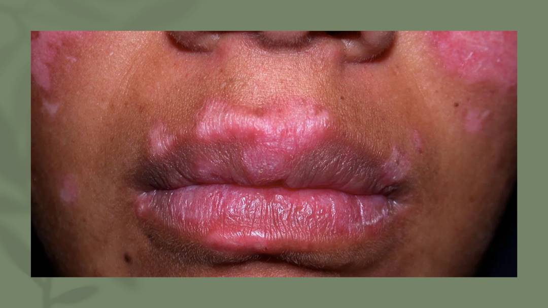
The clinical manifestations of SLE are diverse and can affect multiple organ systems in the body. Common symptoms include fatigue, fever, joint pain and swelling, skin rashes, and photosensitivity. SLE can also cause more serious complications such as lupus nephritis, which is a major cause of morbidity and mortality in patients with SLE.
ASSESSMENT AND DIAGNOSTIC FINDINGS
The diagnosis of SLE is based on a combination of clinical and laboratory findings. The American College of Rheumatology has developed diagnostic criteria for SLE, which require the presence of at least four of the following: malar rash, discoid rash, photosensitivity, oral ulcers, arthritis, serositis, renal disorder, neurologic disorder, hematologic disorder, immunologic disorder, and antinuclear antibody positivity. Laboratory tests that may be helpful in diagnosing SLE include antinuclear antibody (ANA) testing, anti-double-stranded DNA (anti-dsDNA) antibody testing, and complement-level testing.
MEDICAL MANAGEMENT
The management of SLE involves a multidisciplinary approach, including rheumatologists, nephrologists, dermatologists, and other specialists as needed. Treatment goals include controlling disease activity, preventing flares, and minimizing organ damage. Treatment options may include nonsteroidal anti-inflammatory drugs (NSAIDs), antimalarial drugs, glucocorticoids, immunosuppressants, and biological agents.
PHARMACOLOGIC MANAGEMENT
Pharmacologic management of SLE involves a range of medications targeting different aspects of the disease’s pathophysiology. NSAIDs can be used to manage mild to moderate pain and inflammation, while antimalarial drugs such as hydroxychloroquine can be used to prevent disease flares and reduce disease activity. Glucocorticoids such as prednisone can be used to manage severe disease activity and organ involvement, but their long-term use is associated with significant adverse effects. Immunosuppressive agents such as azathioprine, mycophenolate mofetil, and cyclophosphamide can be used to control disease activity and prevent organ damage. Biologic agents such as belimumab, a monoclonal antibody targeting B-cell activating factor, have also been approved for the treatment of SLE.
Systemic lupus erythematosus diagnosis and management, https://academic.oup.com/rheumatology/article/56/suppl_1/i3/2738661.
C. (2023, January 31). Systemic lupuserythematosus (SLE). Centers for Disease Control and Prevention. https://www.cdc.gov/lupus/facts/detailed.html
Systemic lupus erythematosus pathophysiology – wikidoc. (n.d.). Systemic Lupus Erythematosus Pathophysiology – Wikidoc. https://www.wikidoc.org/index.php/Systemic_lupus_erythematosus_pathophysiology
Discover more from Nursing In Ghana
Subscribe to get the latest posts sent to your email.
Shock is a threatening life condition of circulatory failure which causes inadequate oxygen delivery to meet cellular metabolic needs and oxygen consumption requirements, producing cellular and tissue hypoxia. The effects of shock are initially reversible, but rapidly become irreversible, resulting in multiorgan failure (MOF) and death. When a patient presents with undifferentiated shock, it is important that the clinician immediately initiate therapy while rapidly identifying the etiology so that definitive therapy can be administered to reverse shock and prevent MOF and death.
There are four main types of shock:
1. Anaphylactic shock
2. Cardiogenic shock
3. Hypovolemic shock
4. Septic shock
Anaphylactic shock is a severe and sudden allergic reaction that can occur after exposure to an allergen. Symptoms include swelling of the face and throat, difficulty breathing, and a drop in blood pressure. Anaphylactic shock can be life-threatening and requires immediate medical treatment.
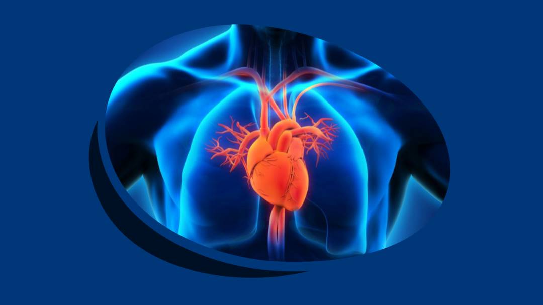
Cardiogenic shock occurs when the heart is unable to pump enough blood to meet the body’s needs. This can be due to a heart attack, heart failure, or other heart conditions. Symptoms include shortness of breath, chest pain, and a weak and irregular heartbeat. Cardiogenic shock is a medical emergency and requires treatment in a hospital.
Hypovolemic shock occurs when there is a decrease in the amount of blood or fluid in the body. This can be due to blood loss from an injury, severe dehydration, or excessive vomiting or diarrhea. Symptoms include lightheadedness, fainting, and a decrease in urine output. Hypovolemic shock can be life-threatening and requires immediate medical treatment.
Septic shock. This type of shock is caused by an infection or sepsis. Sepsis is a potentially life-threatening condition that occurs when an infection spreads throughout the body. Symptoms include low blood pressure, rapid heart rate, chills, and fever. Septic shock is a medical emergency and requires treatment in a hospital.
Discover more from Nursing In Ghana
Subscribe to get the latest posts sent to your email.

FUNERAL HELD FOR GHANAIAN MIDWIFE, GRACE BENYIN WHO DIED IN IRELAND

GHANA, GRENADA SIGN NURSE RECRUITMENT DEAL – APPLICATIONS OPEN OCT 24-28, 2025

Scotland NHS Recruiting International Nurses
Ministry of Health Postpones Media Engagement on Unpaid Salaries

NMC Issues Strong Warning to Nurses Against Unprofessional Conduct on Social Media
Government slashes 50% off Nursing Training Fees-Press Release

GRNMA Youth Committee launches 2025 International Youth Day celebration
President Mahama promises UHAS expansion to train more health workers
LIST OF ACCREDITED GOVERNMENT NURSING AND MIDWIFERY TRAINING SCHOOLS IN GHANA

NURSING ADMISSION FORMS ON SALE FOR THE 2023/2024 ACADEMIC YEAR

THE NURSES PLEDGE AND THE MIDWIVE’S PRAYER

NURSING TRAINING ADMISSION INTERVIEW QUESTIONS

GHS INTRODUCES TWO NEW BELT COLOURS FOR TWO NEW LEVELS IN THE NURSING AND MIDWIFERY SERVICE

MOH SUSPENDS THE 2021/2022 ACADEMIC CALENDAR FOR NURSING AND MIDWIFERY SCHOOLS

COMMON TYPES OF INTRAVENOUS (IV) FLUIDS AND THEIR USES

LIST OF PRIVATE NURSING AND MIDWIFERY TRAINING SCHOOLS (ACCREDITED)
-
 Nursing News5 years ago
LIST OF ACCREDITED GOVERNMENT NURSING AND MIDWIFERY TRAINING SCHOOLS IN GHANA
-
 Nursing News3 years ago
NURSING ADMISSION FORMS ON SALE FOR THE 2023/2024 ACADEMIC YEAR
-
 Nursing Procedures and Skills5 years ago
THE NURSES PLEDGE AND THE MIDWIVE’S PRAYER
-
 Nursing Procedures and Skills5 years ago
NURSING TRAINING ADMISSION INTERVIEW QUESTIONS
-
 Nursing News4 years ago
GHS INTRODUCES TWO NEW BELT COLOURS FOR TWO NEW LEVELS IN THE NURSING AND MIDWIFERY SERVICE
-
 Nursing News5 years ago
MOH SUSPENDS THE 2021/2022 ACADEMIC CALENDAR FOR NURSING AND MIDWIFERY SCHOOLS
-
 Notes5 years ago
COMMON TYPES OF INTRAVENOUS (IV) FLUIDS AND THEIR USES
-
 Nursing News5 years ago
LIST OF PRIVATE NURSING AND MIDWIFERY TRAINING SCHOOLS (ACCREDITED)